Regeneration (biology)


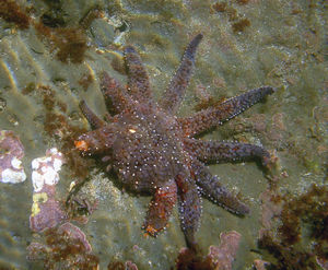
In biology, an organism is said to regenerate a lost or damaged part if the part regrows so that the original function is restored. Regenerative capacity is inversely related to complexity: in general, the more complex an animal is the less regeneration it is capable of. Whereas newts, for example, can regenerate severed limbs, mammals cannot. Limb regeneration in newts occurs in two major steps, first de-differentiation of adult cells into a stem cell state similar to embryonic cells and second, development of these cells into new tissue more or less the same way it developed the first time.[1] Simpler animals like planarian have an enhanced capacity to regenerate because the adults retain clusters of stem cells within their bodies which migrate to the parts of the body that need healing then divide and differentiate to provide the required missing tissue.
Contents |
Regeneration in amphibians
In salamanders, the regeneration process begins immediately after amputation. Limb regeneration in the axolotl and newt have been extensively studied. After amputation, the epidermis migrates to cover the stump in less than 12 hours, forming a structure called the apical epidermal cap (AEC). Over the next several days there are changes in the underlying stump tissues that result in the formation of a blastema (a mass of dedifferentiated proliferating cells). As the blastema forms, pattern formation genes – such as HoxA and HoxD – are activated as they were when the limb was formed in the embryo.[2][3] The distal tip of the limb (the autopod, which is the hand or foot) is formed first in the blastema. The intermediate portions of the pattern are filled in during growth of the blastema by the process of intercalation.[1][2] Motor neurons, muscle, and blood vessels grow with the regenerated limb, and reestablish the connections that were present prior to amputation. The time that this entire process takes varies according to the age of the animal, ranging from about a month to around three months in the adult and then the limb becomes fully functional.
In spite of the historically small size of the number of researchers studying limb regeneration, remarkable progress has been made recently in establishing the neotenous amphibian the axolotl (Ambystoma mexicanum) as a model genetic organism. This progress has been facilitated by advances in genomics, bioinformatics, and somatic cell transgenesis in other fields, that have created the opportunity to investigate the mechanisms of important biological properties, such as limb regeneration, in the axolotl.[4] The Ambystoma Genetic Stock Center (AGSC) is a self-sustaining, breeding colony of the axolotl supported by the National Science Foundation as a Living Stock Collection. Located at the University of Kentucky, the AGSC is dedicated to supplying genetically well-characterized axolotl embryos, larvae, and adults to laboratories throughout the United States and abroad. An NIH-funded NCRR grant has led to the establishment of the Ambystoma EST database, the Salamander Genome Project (SGP) that has led to the creation of the first amphibian gene map and several annotated molecular data bases, and the creation of the research community web portal.[5]
Regeneration in Planaria
Planaria exhibit an extraordinary ability to regenerate lost body parts. For example, a planarian split lengthwise or crosswise will regenerate into two separate individuals. In one experiment, T. H. Morgan found that a piece corresponding to 1⁄ 279th of a planarian could successfully regenerate into a new worm. This size (about 10,000 cells) is typically accepted as the smallest fragment that can regrow into a new planarian.
Regeneration of human skeleton
Finger Tips
Studies in the 1970s showed that children up to the age of 10 or so who lose fingertips in accidents can regrow the tip of the digit within a month provided their wounds are not sealed up with flaps of skin – the de facto treatment in such emergencies. They normally won't have a finger print, and if there is any piece of the finger nail left it will grow back as well, usually in a square shape rather than round.[6][7]
In August 2005, Lee Spievack, then in his early sixties, accidentally sliced off the tip of his right middle finger just above the first phalanx. His brother, Dr. Alan Spievack, was researching regeneration and provided him with powdered extracellular matrix, developed by Dr. Stephen Badylak of the McGowan Institute of Regenerative Medicine. Mr. Spievack covered the wound with the powder, and the tip of his finger re-grew in four weeks.[8] The news was released in 2007. Lee Spievack is the first documented case of an adult human regenerating fingertips;[6] however, Ben Goldacre has described this as "the missing finger that never was", claiming that fingertips regrow and quoted Simon Kay, professor of hand surgery at the University of Leeds, who from the picture provided by Goldacre described the case as seemingly "an ordinary fingertip injury with quite unremarkable healing" and as "junk science".[9]
Ribs
There have appeared claims that human ribs could regenerate if the periosteum, the membrane surrounding the rib, were left intact. In one study rib material was used for skull reconstruction and all 12 patients had complete regeneration of the resected rib.[10]
Regeneration of human liver
The human liver is one of the few glands in the body that has the ability to regenerate from as little as 25% of its tissue.[11] This is largely due to the unipotency of hepatocytes.[12] Resection of liver can induce the proliferation of the remained hepatocytes until the lost mass is restored, where the intensity of the liver’s response is directly proportional to the mass resected. For almost 80 years surgical resection of the liver in rodents has been a very useful model to the study of cell proliferation.[13][14]
Kidney regeneration
Regenerative capacity of the kidney remains largely unexplored. The basic functional and structural unit of the kidney is nephron, which is mainly composed of four components: the glomerulus, tubules, the collecting duct and peritubular capillaries. The regenerative capacity of the mammalian kidney is limited compared to that of lower vertebrates.
Regeneration in the mammalian kidney
In the mammalian kidney, the regeneration of the tubular component following an acute injury is well known. Recently regeneration of the glomerulus has also been documented. Following an acute injury, the proximal tubule is damaged more, and the injured epithelial cells slough off the basement membrane of the nephron. The surviving epithelial cells, however, undergo migration, dedifferentiation, proliferation, and redifferentiation to replenish the epithelial lining of the proximal tubule after injury. Recently, the presence and participation of kidney stem cells in the tubular regeneration has been shown. However, the concept of kidney stem cells is currently emerging. In addition to the surviving tubular epithelial cells and kidney stem cells, the bone marrow stem cells have also been shown to participate in regeneration of the proximal tubule, however, the mechanisms remain controversial. Recently, studies examining the capacity of bone marrow stem cells to differentiate into renal cells are emerging [15].
Regeneration in the lower vertebrate kidney
Like other organs, the kidney is also known to regenerate completely in lower vertebrates such as fish. Some of the known fish that show remarkable capacity of kidney regeneration are goldfish, skates, rays, and sharks. In these fish, the entire nephron regenerates following injury or partial removal of the kidney.
Regeneration in MRL mice
The mechanism for regeneration in MRL mice has been found and it is related to the deactivation of the p21 gene.[16][17]
Adult mammals have limited regenerative capacity compared to most vertebrate embryos/larvae, adult salamanders and fish. The MRL mouse is a strain of mouse that exhibits remarkable regenerative abilities for a mammal. Study of the regenerative process in these animals is aimed at discovering how to duplicate them in humans.
By comparing the differential gene expression of scarless healing MRL mice and poor healing C57BL/6 mice strain, 36 genes have been identified that are good candidates for studying how the healing process differs in MRL mice and other mice.[18][19]
The regenerative abilities of MRL mice does not, however, protect them against myocardial infarction, as heart regeneration in adult mammals (neocardiogenesis) is limited because heart muscle cells are nearly all terminally differentiated. MRL mice show the same amount of cardiac injury and scar formation as normal mice after a heart attack.[20] Though recent studies provide evidence that this may not be the case, and that MRL mice do regenerate from heart damage. [1]
Notes
- ↑ 1.0 1.1 Odelberg SJ.Unraveling the molecular basis for regenerative cellular plasticity.PLoS Biol. 2004 Aug;2(8):E232. PMID 15314652
- ↑ 2.0 2.1 Bryant, S.V., Endo, T. and Gardiner, D.M. Vertebrate limb regeneration and the origin of limb stem cells. Int. J. Dev. Biol. 2002 46:887-896. PMID 12455626
- ↑ Mullen LM, Bryant SV, Torok MA, Blumberg B, Gardiner DM. Nerve dependency of regeneration: the role of Distal-less and FGF signaling in amphibian limb regeneration. Development. 1996 Nov;122(11):3487-97. PMID 8951064
- ↑ Endo T, Bryant SV, Gardiner DM. A stepwise model system for limb regeneration. Dev Biol. 2004 Jun 1;270(1):135-45. PMID 15136146
- ↑ http://www.ambystoma.org
- ↑ 6.0 6.1 Weintraub, Arlene (MAY 24, 2004). "The Geniuses Of Regeneration". BusinessWeek. http://www.businessweek.com/magazine/content/04_21/b3884008_mz001.htm.
- ↑ Illingworth, Cynthia M. 1974. Trapped fingers and amputated fingertips in children. J. Ped. Surgery 9:853-858.
- ↑ "Regeneration recipe: Pinch of pig, cell of lizard". Associated Press. MSNBC. February 19, 2007. http://www.msnbc.msn.com/id/17171083/. Retrieved October 24, 2008.
- ↑ Goldacre, Ben (May 3, 2008). "The missing finger that never was". The Guardian. http://www.guardian.co.uk/science/2008/may/03/medicalresearch.health.
- ↑ Munro IR, Guyuron B (November 1981). "Split-Rib Cranioplasty". Annals of Plastic Surgery 7 (5): 341–346. doi:10.1097/00000637-198111000-00001. PMID 7332200. PMID 7332200.
- ↑ "Liver Regeneration Unplugged". Bio-Medicine. 2007-04-17. http://www.bio-medicine.org/medicine-news/Liver-Regeneration-Unplugged-19988-1/. Retrieved 2007-04-17.
- ↑ Michael, Dr. Sandra Rose (2007). "Bio-Scalar Technology: Regeneration and Optimization of the Body-Mind Homeostasis" (PDF). 15th Annual AAAAM Conference: 2. http://eesystem.com/docs/AAAAM%202007%20long%20biography%20abstr_.pdf. Retrieved October 24, 2008.
- ↑ Higgins, GM and RM Anderson RM (1931). "Experimental pathology of the liver. I. Restoration of the liver of the white rat following partial surgical removal". Arch. Pathol. 12: 186–202.
- ↑ Michalopoulos, GK and MC DeFrances (April 4, 1997). "Liver regeneration". Science 276 (5309): 60–66. doi:10.1126/science.276.5309.60. PMID 9082986. PMID 9082986.
- ↑ Kurinji Singaravelu et al.(July 2009). "In Vitro Differentiation of MSC into Cells with a Renal Tubular Epithelial-Like Phenotype". Renal Failure 31(6):492-502. http://www.informaworld.com/smpp/content~content=a913452182~db=all~jumptype=rss
- ↑ Bedelbaeva K, Snyder A, Gourevitch D, Clark L, Zhang X-M, Leferovich J, Cheverud JM, Lieberman P, Heber-Katz E (March 2010). "Lack of p21 expression links cell cycle control and appendage regeneration in mice". Proceedings of the National Academy of Sciences 107 (11): 5845–50. doi:10.1073/pnas.1000830107. PMID 20231440. PMC 2851923. http://www.pnas.org/content/early/2010/03/08/1000830107.abstract. Lay summary – PhysOrg.com.
- ↑ Humans Could Regenerate Tissue Like Newts By Switching Off a Single Gene
- ↑ Biochem Biophys Res Commun. 2005 Apr 29;330(1):117-22. PMID 15781240
- ↑ Mansuo L. Hayashi, B. S. Shankaranarayana Rao, Jin-Soo Seo, Han-Saem Choi, Bridget M. Dolan, Se-Young Choi, Sumantra Chattarji, and Susumu Tonegawa (2007 July). "Inhibition of p21-activated kinase rescues symptoms of fragile X syndrome in mice". Proceedings of the National Academy of Sciences 104 (27): 11489. doi:10.1073/pnas.0705003104. PMID 17592139.
- ↑ Abdullah I, Lepore JJ, Epstein JA, Parmacek MS, Gruber PJ (2005 Mar-April). "MRL mice fail to heal the heart in response to ischemia-reperfusion injury.". Wound Repair Regen 13 (2): 205–208. doi:10.1111/j.1067-1927.2005.130212.x. PMID 15828946. PMID 15828946.
Sources
- Tanaka EM. Cell differentiation and cell fate during urodele tail and limb regeneration. Curr Opin Genet Dev. 2003 Oct;13(5):497-501. PMID 14550415
- Nye HL, Cameron JA, Chernoff EA, Stocum DL. Regeneration of the urodele limb: a review. Dev Dyn. 2003 Feb;226(2):280-94. PMID 12557206
- Yu H, Mohan S, Masinde GL, Baylink DJ. Mapping the dominant wound healing and soft tissue regeneration QTL in MRL x CAST. Mamm Genome. 2005 Dec;16(12):918-24. PMID 16341671
- Gardiner DM, Blumberg B, Komine Y, Bryant SV. Regulation of HoxA expression in developing and regenerating axolotl limbs. Development. 1995 Jun;121(6):1731-41. PMID 7600989
- Torok MA, Gardiner DM, Shubin NH, Bryant SV. Expression of HoxD genes in developing and regenerating axolotl limbs. Dev Biol. 1998 Aug 15;200(2):225-33. PMID 9705229
- Putta S, Smith JJ, Walker JA, Rondet M, Weisrock DW, Monaghan J, Samuels AK, Kump K, King DC, Maness NJ, Habermann B, Tanaka E, Bryant SV, Gardiner DM, Parichy DM, Voss SR, From biomedicine to natural history research: EST resources for ambystomatid salamanders. BMC Genomics. 2004 Aug 13;5(1):54. PMID 15310388
- Andrews, Wyatt (March 23, 2008). "Medicine's Cutting Edge: Re-Growing Organs". Sunday Morning (CBS News). http://www.cbsnews.com/stories/2008/03/22/sunday/main3960219.shtml.
External links
- Spallanzani's mouse: a model of restoration and regeneration
- Mice that regrow hearts in the news
- DARPA Grant Supports Research Toward Realizing Tissue Regeneration
- The Geniuses Of Regeneration in BusinessWeek, May 24, 2004
- UCI Limb Regeneration Lab
|
||||||||